
Pet Ultrasound in Lehi, UT
Better Care for Your Pet
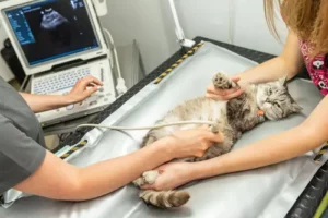
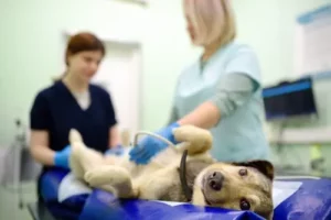
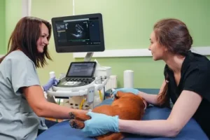
When a pet is sick and the cause is not obvious, clear diagnostic information can make all the difference. At Lehi Hills Veterinary Hospital, we provide pet ultrasound in Lehi, UT, to help our veterinary team examine the internal organs of dogs and cats in real time. Ultrasound uses sound waves and returning echoes to create images of the abdomen without surgery. Combined with our pet imaging,digital radiography, physical examinations, and in-hospital lab work, this technology helps us investigate symptoms, confirm findings, and guide thoughtful treatment decisions for your pet.
What a Pet Ultrasound Can Show
An ultrasound gives us a live view of structures inside your pet’s abdomen. Unlike an X-ray, which is especially useful for evaluating bones, gas patterns, and the general shape of organs, ultrasound allows us to see the texture, movement, and internal appearance of many soft tissues.
Depending on your pet’s symptoms, we may use ultrasound to evaluate:
- The liver and gallbladder
- Kidneys and urinary bladder
- Spleen
- Stomach and intestines
- Pancreas
- Adrenal glands
- Reproductive organs
- Abdominal lymph nodes
- Abnormal fluid or masses
Because the images appear in real time, we can observe movement within the abdomen and examine suspicious areas from different angles.
When We May Recommend an Ultrasound
Pets cannot tell us exactly where they hurt or what feels different. Symptoms such as vomiting, diarrhea, weight loss, appetite changes, or abdominal swelling may have several possible causes. Diagnostic imaging helps us narrow those possibilities and determine the next appropriate service for your furry friend.
We may recommend a pet ultrasound when your dog or cat has:
- Abnormal blood work results
- Vomiting or diarrhea that is not responding to treatment
- A distended or painful abdomen
- An irregular finding during abdominal palpation
- Unexplained weight loss
- Reduced appetite
- Changes in urination
- Suspected internal fluid
- An abnormality identified on an X-ray
- Symptoms that continue without a clear diagnosis
Ultrasound may also help us monitor a known condition or evaluate how an organ has changed over time.
Confirming Findings From Digital Radiography
Digital radiography and ultrasound often work together. X-rays provide a broad view of the abdomen and may reveal changes in organ size, unusual gas patterns, mineral material, foreign objects, or other abnormalities. Ultrasound can then help us look more closely at the affected soft tissues.
For example, an X-ray may show that an organ appears enlarged or that something unusual is present in the abdomen. Ultrasound can provide additional detail about the organ’s internal structure and surrounding tissues.
Our access to digital radiography for pets allows us to review clear images efficiently and decide whether further pet imaging is needed.
In-Hospital Lab Work for Dogs and Cats
Imaging is only one part of a complete diagnostic evaluation. Blood tests, urine testing, and other laboratory results can provide important information about organ function, infection, inflammation, hydration, and blood cell levels.
At Lehi Hills Veterinary Hospital, we offer in-hospital lab work for dogs and cats. Having access to laboratory testing within our hospital helps us connect your pet’s symptoms with imaging findings more quickly.
For instance, abnormal liver values may lead us to examine the liver and gallbladder more closely. Kidney changes in blood work may make a detailed evaluation of the kidneys and bladder appropriate. By combining these tools, we gain a more complete understanding of your pet’s health.
Collecting Cytology Samples With Ultrasound Guidance
In certain cases, ultrasound can help guide the collection of cytologic samples from an abnormal area. Cytology involves examining cells under a microscope to gather more information about a mass, organ, fluid pocket, or other finding.
Ultrasound guidance allows us to see the target area while collecting the sample, which can improve accuracy and help us avoid nearby structures. The collected material may be evaluated in-house or sent to a laboratory, depending on the type of sample and the information needed.
Not every abnormality requires sampling. We discuss the potential benefits, limitations, and next steps with you before recommending this procedure.
What to Expect During Your Pet’s Ultrasound
Before the appointment, our team will explain whether your pet needs to avoid food or follow other preparation instructions. In many cases, we clip a small area of fur over the abdomen so the ultrasound probe can make proper contact with the skin.
A clear gel is applied to the area, and the probe is moved gently across the abdomen to create images. Most pets can remain awake during the examination, although mild sedation may be recommended if a pet is anxious, painful, or unable to remain still.
We focus on keeping the experience as calm and comfortable as possible. Afterward, we explain the findings and discuss whether treatment, monitoring, laboratory testing, or additional diagnostics are recommended.
A More Complete Approach to Veterinary Diagnosis
Ultrasound does not replace every other diagnostic tool. Instead, it adds valuable information to the overall picture. Physical examination findings, medical history, lab work, digital radiography, and ultrasound each answer different questions.
Our veterinary team uses these tools together to avoid making decisions based on a single symptom or test. We want to understand not only what is happening, but also how the findings relate to your pet’s comfort, daily behavior, and overall health.
This comprehensive approach helps us create a treatment plan based on useful evidence rather than guesswork.
Frequently Asked Questions
No. Ultrasound is generally noninvasive and painless. Your pet may feel gentle pressure from the probe, but most dogs and cats tolerate the examination well.
Many pets do not require sedation. It may be recommended when a pet is very nervous, painful, or unable to remain still long enough for clear images.
Clipping allows the ultrasound probe to contact the skin directly. Fur can trap air and interfere with the sound waves needed to produce a clear image.
The length of the appointment depends on the area being evaluated, your pet’s comfort, and the complexity of the findings. Our team can provide more specific expectations before the exam.
No single test can identify every condition. Ultrasound provides detailed information about many soft tissues, but lab work, X-rays, cytology, or other testing may also be needed.
We discuss available findings after the examination. If samples are sent to an outside laboratory or additional review is needed, those results may take longer.
Get Clearer Answers About Your Pet’s Health
Unexplained symptoms can be stressful, especially when your pet is uncomfortable and you do not yet know why. Our ultrasound, digital radiography, and in-hospital laboratory capabilities help us investigate concerns efficiently and build a more complete picture of your pet’s condition. At Lehi Hills Veterinary Hospital, we combine modern diagnostic tools with attentive, compassionate care for dogs and cats throughout Lehi and nearby Utah communities. Contact our vets today to schedule an evaluation and learn whether ultrasound may help provide the answers your pet needs.